NT Scan in Pregnancy: Nuchal Translucency Test, Procedure & Report
Published on: Jun 10, 2026
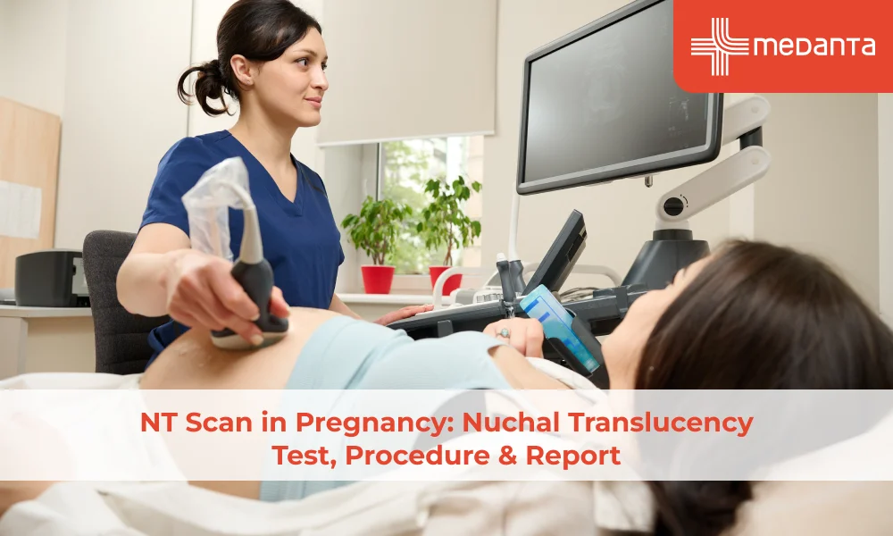
TABLE OF CONTENTS
Overview of NT Scan
The first trimester offers a narrow but important window for early screening. The NT scan measures a fluid-filled space at the back of the baby's neck and combined with a blood test and maternal age apriori risk produces a personalised risk estimate for chromosomal conditions. Understanding what it does and equally what it does not helps parents engage with results without unnecessary anxiety.
What Is an NT Scan?
The nuchal translucency (NT) scan is a first-trimester ultrasound that measures the nuchal translucency which is the fluid-filled space (translucency) at the back of the foetal neck (nuchal means relating to the neck). In a normal pregnancy, all foetuses have a small amount of fluid in this space. The key finding is how much. Fluid accumulates in this area when foetal lymphatic drainage is immature or obstructed, which is more common in foetuses with chromosomal abnormalities or structural defects especially cardiac.
The NT scan is interpreted alongside with a maternal blood test measuring two specific markers - free beta-human chorionic gonadotrophin (free beta-hCG, a hormone produced by the placenta) and pregnancy-associated plasma protein A (PAPP-A, a protein produced by the developing placenta). Together, the NT measurement, the blood markers and the mother's age produce a combined risk calculation. This combined first-trimester screening is more accurate than any single component alone.
Why and When an NT Scan Is Done During Pregnancy
The NT scan is offered between 11 weeks and 13 weeks 6 days when the CRL is 45–84 mm. Before 11 weeks the NT space is too small to measure and after 14 weeks the fluid is absorbed and the measurement loses meaning. Its purpose is screening, not diagnosis as a high-risk result means elevated probability, not certainty and further testing is warranted. It screens most reliably for:
• Trisomy 21 (Down syndrome): Caused by an extra copy of chromosome 21
• Trisomy 18 (Edwards syndrome): A severe condition from an extra chromosome 18
• Trisomy 13 (Patau syndrome): A severe condition from an extra chromosome 13
• Turner syndrome and other sex chromosome conditions
• Major structural heart defects: A raised NT is a known marker for congenital heart disease even when chromosomes are normal.
How the NT Scan Procedure Is Performed
The NT scan is a standard abdominal ultrasound: gel is applied and a transducer transmits sound waves that produce real-time images. No radiation is involved and the scan is painless. Achieving the correct NT measurement is technically demanding - the baby must lie in profile with the head and body aligned (a sagittal midline view). The mother may be asked to walk or roll to encourage the right position. A transvaginal ultrasound is occasionally needed and is offered only with the mother's consent.
Understanding NT Scan Results and Normal Range
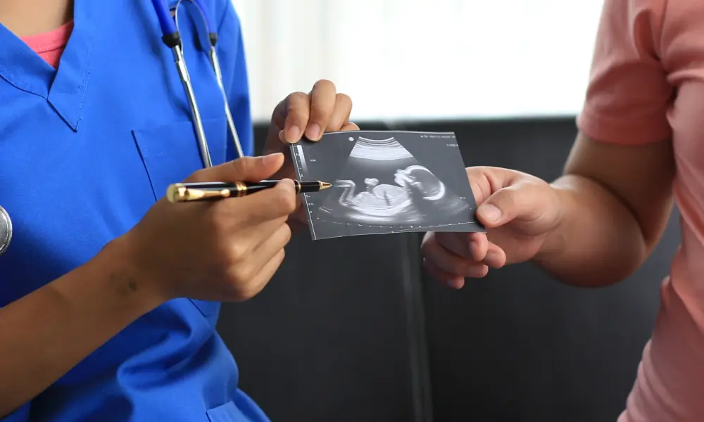
The NT measurement is reported in millimetres. What is considered "normal" depends on crown-rump length as the NT space grows slightly as the baby grows, so it is always interpreted relative to the baby's size. As a general guide:
• NT below 2.5 mm at 11–14 weeks is generally considered within the normal range for most gestational ages
• NT of 3.5 mm or above is associated with a significantly higher risk of chromosomal abnormality or structural heart defect
• NT at or above 3.5 mm with a normal chromosomal result still needs a detailed foetal cardiac scan (fetal echocardiography) at 20–22 weeks.
These thresholds are guides, not cut-offs. The combined risk calculation incorporating NT, PAPP-A, free beta-hCG, and maternal age produces a single risk figure.
Risks and Limitations of the NT Scan
Non-invasive and carrying no physical risk, the NT scan's principal limitation is accuracy as a screening test:
• Detection rate: NT scan alone has sensitivity of ~ 70% but when we do first trimester combined screening sensitivity increases up to 87-89% of Down syndrome cases detected at a 5% false-positive rate meaning roughly 1 in 20 women receive a high-risk result that turns out, on diagnostic testing, to be unaffected
• False negatives: Some chromosomal abnormalities are not detected and a low-risk result does not guarantee a normal outcome
• Operator dependency: NT measurement requires certified sonographers and the quality varies between centres
• Limited scope: the test does not screen for single-gene disorders, submicroscopic chromosomal changes, or most structural abnormalities - these require separate testing.
What Happens if the NT Scan Result Is Abnormal?
A high-risk result leads to a discussion with a fetal medicine specialist. Options are:
• Non-invasive prenatal testing (NIPT): a blood test analysing foetal DNA fragments in the mother's blood, detecting ~98% of Down syndrome cases. Although noninvasive screening test but sensitivity is not same for all chromosomes and a positive test result must be confirmed by an invasive test before decisions are made.
• Chorionic villus sampling (CVS): a small placental tissue sample taken at 11–14 weeks and chromosomes are examined directly, providing a definitive result. .
• Amniocentesis: amniotic fluid is sampled at 15–20 weeks, also providing a definitive chromosomal diagnosis.
Both invasive tests provide certainty that no screening test can.
FAQs
Is the NT scan mandatory during pregnancy?
In India, it forms part of the standard first-trimester pathway recommended by FOGSI, but participation is voluntary. Some women decline because they would not act on any result. The purpose is informed decision-making, not compulsion.
2. Can the NT scan detect all genetic conditions?
No. The combined test detects common trisomies like Down syndrome (trisomy 21), Edwards syndrome (trisomy 18) and patau syndrome (trisomy 13) It does not detect other chromosomal or single-gene disorders, submicroscopic chromosomal changes or most structural abnormalities. A normal result does not exclude all genetic conditions.
3.Is the NT scan painful or invasive?
The standard NT scan is an abdominal ultrasound that is entirely non-invasive and painless. If a transvaginal ultrasound (probe inserted into the vagina) is needed for a clearer view, it is offered with full explanation and separate consent and most women find it briefly uncomfortable rather than painful. The scan carries no risk to the pregnancy.
4. How accurate is the NT scan in pregnancy screening?
As a standalone measurement, NT detects the risk of chromosomal conditions like Down syndrome. Its accuracy improves significantly when combined with PAPP-A, free beta-hCG, and maternal age. No screening test is perfect, but the combined test is among the most effective first-trimester tools available.
5. What happens if the NT scan shows a higher-than-normal measurement?
A raised NT is discussed with a fetal medicine specialist. For a definitive diagnosis doctors suggest further testing e.g invasive testing(CVS or amniocentesis)
Can the NT scan determine the baby's gender?
India's Pre-Conception and Prenatal Diagnostic Techniques (PCPNDT) Act 1994 prohibits disclosure of foetal sex during prenatal scans.
7. Is any special preparation needed before an NT scan?
Drink 500–750 mL of water 30–45 minutes beforehand as a partially filled bladder lifts the uterus, improving the view. No fasting is required. Wear loose clothing for easy abdominal access.
8. Can an NT scan be done after the first trimester?
No the NT space is only measurable between 11 and 13 weeks 6 days when the CRL is 45–84 mm. After 14 weeks the lymphatic system matures, the fluid is absorbed, and measurement is no longer possible. Women who miss this window can have second-trimester screening (quadruple screening test) , though these are less sensitive or NIPT.
9. Are additional tests required after an abnormal NT scan report?
In case of an abnormal NT scan, doctors recommend further testing NIPT or invasive diagnostic testing like CVS or amniocentesis. The appropriate step depends on risk level, type of abnormality,gestational age, and the couple's preferences.
10. Is the NT scan safe for both mother and baby?
Yes diagnostic ultrasound at routine obstetric power levels has been used for over 50 years without evidence of harm. No radiation is involved and no long-term adverse effects have been identified. It is among the safest diagnostic tools in pregnancy.