1068
1068
 Request Call Back
 Contact
 1068
1068


Largest team of surgeons specialising in minimally invasive techniques such as Video-assisted Thoracic Surgery (VATS) and Robot-assisted Thoracic Surgery (RATS) with exceptional success rate, low mortality, morbidity, and complications

Enquiry Form

Search for Hospitals
Chest Surgery Highlights

01
One of India's Largest and Most Experienced Centres for Chest Surgery
02
Comprehensive Care for Every Disease of the Chest — From Diagnosis to Recovery
03
Cutting edge surgical care for all diseases of Chest, including cancers.
04
Over 5000 major Chest surgeries and 1000+ Robotic Chest Surgeries in India
05
Expertise in complex airway surgeries with recurrence rate on a par with best centre in the world.
06
High-Volume thoracic surgery centre using Minimally Invasive VATS /Robotic Techniques.
About the Speciality
Medanta Institute of Chest Surgery and Chest-Onco Surgery is a specialized centre dedicated to the comprehensive management of all diseases and disorders of the chest (thoracic cavity), including thoracic cancers. Through a multidisciplinary and multimodal approach, we address the entire spectrum of patient care—from prevention and early intervention to advanced medical, interventional, and surgical treatments. Our team comprises dedicated specialists with expertise in diseases of the lungs, trachea and bronchi, pleura, mediastinum, oesophagus, chest wall, and diaphragm, supported by nurses specially trained in thoracic care. We utilize advanced diagnostic technologies to accurately assess the stage and complexity of each condition, enabling us to develop personalized treatment and recovery plans for every patient. Our chest surgeons, thoracic oncology surgeons, and lung transplant experts work collaboratively with specialists across disciplines to deliver precise, evidence-based care. The institute is at the forefront of minimally invasive thoracic surgery, offering advanced procedures including Video-Assisted Thoracoscopic Surgery (VATS) and robotic-assisted chest surgery, ensuring better outcomes, faster recovery, and enhanced patient comfort.
Our Chairman Message
The rising incidence of chest diseases has necessitated a specialised centre offering the complete range of wellness solutions from fast, accurate, expert diagnosis to holistic and personalised treatments, post surgery rehabilitation and facility for long term follow-up. Medanta Institute of Chest Surgery and Chest-Onco Surgery is dedicated to fulfilling this crucial role. No matter the state or complexity of the disease, our handpicked team of experts – carrying over 10 decades of experience – determines the best course of action for patients. Our endeavour to help everyone breathe better is supported by all super-specialities, including radiation oncology, pulmonary medicine, critical care, cardiac care.

Chest Surgery Team
Unlocking Vitality, Medanta's Mastery in Chest Surgery


State-of-the-art diagnostic facility for cancer screening. Fully modular state-of-the-art operating rooms with world-class facility dedicated to thoracic surgery. Use of advanced surgical robots for high precision, reduced side effects and faster recovery.

End-to-end comprehensive care, including disease prevention, reversal, clinical treatment (interventional and surgical) for all stages of thoracic disease, including cancers. Complete post-surgery support including rehabilitation and nutrition.

Dedicated specialists to treat cancers and some of the most complex and rare diseases of the chest. Our clinicians are supported by a team of nurses specially trained in thoracic disease management

Specialised team to handle all thoracic surgical emergencies, including chest trauma. Expertise in the management of tracheobronchial injury and severe chest wall injuries. Excellent critical care facility with the provision of ECMO.
Our Treatments
Unlocking Vitality through Expert Chest Care.
01
Chest Trauma
Chest trauma encompasses injuries to the chest wall, lungs, and other thoracic structures. Blunt or penetrating trauma can lead to life-threatening conditions, requiring immediate medical attention and, in some cases, surgical intervention.
02
Aspergilloma (Fungus Ball in the Lungs)
An aspergilloma is a dense mass of Aspergillus fungal hyphae, inflammatory cells, and mucus that colonises a pre-existing cavity within the lung (most often a residual cavity left behind by healed tuberculosis, sarcoidosis, or prior abscess). The fungus does not invade lung tissue in immunocompetent individuals; instead it proliferates within the cavity, forming a movable "fungus ball" visible on chest CT as a solid opacity with a surrounding crescent of air (the air-crescent sign). Many patients remain asymptomatic for years, but recurrent or massive haemoptysis which can be caused by erosion of bronchial vessels lining the cavity wall can be severe and occasionally fatal. Antifungal agents like itraconazole or voriconazole have limited penetration into avascular cavities and rarely eradicate established aspergilloma. Surgical resection (most often a lobectomy or segmentectomy) remains the definitive treatment for symptomatic aspergilloma, particularly in patients with good pulmonary reserve, and carries the highest long-term cure rate.
03
Empyema
Empyema is a condition in which pus accumulates in the pleural space—the area between the lungs and the chest wall—usually due to a bacterial infection. It commonly develops as a complication of pneumonia but can also occur after chest surgery or other infections. Symptoms may include fever, chest pain, cough, breathlessness, and fatigue. Treatment typically involves antibiotics and drainage of the infected fluid, with surgery sometimes required in advanced cases.
04
Pneumothorax (Air Around the Lungs)
Pneumothorax occurs when air escapes from the lung and collects in the pleural space (the thin gap between the lung surface and the chest wall) causing partial or complete lung collapse. It can arise spontaneously in young, tall individuals with small subpleural air blebs (primary spontaneous pneumothorax), or as a consequence of underlying lung disease such as COPD, tuberculosis, or cystic fibrosis (secondary spontaneous pneumothorax). Traumatic pneumothorax, caused by penetrating or blunt chest injury, represents a surgical emergency.
Patients typically present with sudden-onset chest pain and breathlessness; in tension pneumothorax, rapid haemodynamic compromise demands immediate needle decompression. First-line management ranges from observation and supplemental oxygen for small, stable episodes to intercostal chest drain insertion for larger collapses. Recurrent or persistent pneumothorax (where the air leak fails to seal spontaneously) requires surgical intervention: VATS pleurodesis or pleurectomy to obliterate the pleural space and prevent further episodes.
05
Myasthenia Gravis and Thymoma
Myasthenia gravis (MG) is a chronic autoimmune disorder in which the body's own immune system attacks the acetylcholine receptors at the neuromuscular junction, disrupting the signal between nerve and muscle. In some MG patients, the thymus gland harbours a tumour called a thymoma (a slow-growing but clinically significant mass that can drive or sustain the autoimmune attack) making the relationship between the two conditions far more than coincidental.
Our Chest Cancer Program
Unlocking Vitality through Expert Chest Care.
Chest Wall Tumours
Chest wall tumours represent a heterogeneous group of neoplasms arising from the bony and cartilaginous skeleton of the thorax (the ribs, sternum, clavicle, and scapula) as well as from soft tissues including muscle, fat, and nerves. They may be primary (originating in the chest wall itself) or secondary (metastatic deposits from breast, prostate, kidney, or lung primaries). Primary bone tumours such as chondrosarcoma, Ewing sarcoma, and osteosarcoma require wide surgical resection with adequate margins to reduce the risk of local recurrence; soft tissue sarcomas of the chest wall are similarly managed with excision guided by oncological principles. Defects created by large resections require immediate chest wall reconstruction to maintain respiratory mechanics and protect intrathoracic organs. At Medanta, this is performed using custom 3D-printed titanium implants and synthetic mesh, planned preoperatively from the patient's own CT data, allowing anatomically precise reconstruction that restores both form and function.
Mediastinal Tumours
The mediastinum - the central chest compartment bounded by the sternum anteriorly, spine posteriorly, and the lungs on either side is divided into anterior, middle, and posterior zones, each with its own characteristic tumour types. Anterior mediastinal masses include thymomas, teratomas and other germ cell tumours, and thyroid or parathyroid lesions. The middle mediastinum is the site of lymphoma and bronchogenic cysts, while the posterior mediastinum is home to neurogenic tumours such as schwannomas and neurofibromas arising from intercostal nerve roots. Clinical presentation depends heavily on size and location with smaller tumours being often silent, while larger ones compress adjacent structures to cause breathlessness, superior vena cava obstruction, Horner's syndrome, or hoarseness from recurrent laryngeal nerve involvement. Tissue characterisation through CT-guided biopsy or endobronchial ultrasound (EBUS) guides treatment planning; surgical resection, feasible by VATS or robotic access in many cases, offers the most complete and durable outcome across the majority of benign and resectable malignant mediastinal tumours.
Lung Cancer
Lung cancer is the leading cause of cancer-related mortality worldwide and the most common malignancy managed by thoracic surgeons. It is broadly classified into non-small cell lung cancer (NSCLC) which includes adenocarcinoma, squamous cell carcinoma, and large cell carcinoma and small cell lung cancer (SCLC), which behaves more aggressively and is treated primarily with chemotherapy and radiotherapy. Adenocarcinoma, arising predominantly in the peripheral lung parenchyma, has become the most prevalent subtype, particularly among non-smokers and women. Symptoms in early-stage disease are often absent or non-specific ranging from a persistent cough, mild breathlessness, or a shadow noticed incidentally on a chest X-ray which explains why most patients still present at an advanced stage. Surgery remains the cornerstone of curative treatment for early and locally advanced NSCLC, with anatomical lung resection (lobectomy or segmentectomy) the standard of care; VATS and robotic lobectomy now allow most patients to undergo this with minimal trauma and faster return to function.
Oesophageal (Food Pipe) Cancer
Oesophageal cancer arises from the mucosal lining of the food pipe and occurs in two principal histological types: squamous cell carcinoma, which predominates in the upper and middle oesophagus and is strongly associated with tobacco use and alcohol, and adenocarcinoma, which develops in the lower oesophagus on a background of chronic gastro-oesophageal reflux disease and Barrett's oesophagus. The cardinal symptom is progressive dysphagia (initially to solids, then liquids) often accompanied by significant weight loss, retrosternal discomfort, and regurgitation. Because the oesophagus has no peritoneal covering and is closely adjacent to major vascular and airway structures, early lymphatic spread is common, and staging must be meticulous. Curative intent treatment for localised disease typically involves neoadjuvant chemoradiotherapy followed by oesophagectomy; Medanta's surgeons perform both open and minimally invasive robotic-assisted oesophagectomy, with the choice guided by tumour location, patient physiology, and oncological staging.
Thymoma
Thymoma is a tumour arising from the epithelial cells of the thymus gland, located in the anterior mediastinum behind the sternum. It is the most common primary tumour of the anterior mediastinum in adults, though it remains relatively rare in absolute terms. Thymomas are graded histologically according to the WHO classification (Types A, AB, B1, B2, B3) - a system that broadly predicts the likelihood of local invasiveness and long-term recurrence. Many patients are asymptomatic, with the tumour discovered incidentally on imaging done for another purpose; others present with chest discomfort, a persistent cough, or superior vena cava syndrome from local compression. A significant proportion are diagnosed because they develop myasthenia gravis (the autoimmune link that makes thymoma unique among thoracic tumours). Complete surgical resection is the primary and most effective treatment, and at Medanta, this is achieved using robotic or VATS thymectomy for early-stage disease, with open resection and HITHOC reserved for locally advanced or pleural-disseminated thymomas.
Latest Technology Utilisation
Our Chest Surgery Department comes fully equipped with:
- HITHOC (Hyperthermic Intrathoracic Chemotherapy) for Advanced Thymomas
- 3D Reconstruction and Implants for Chest Wall Tumours
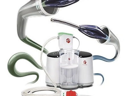
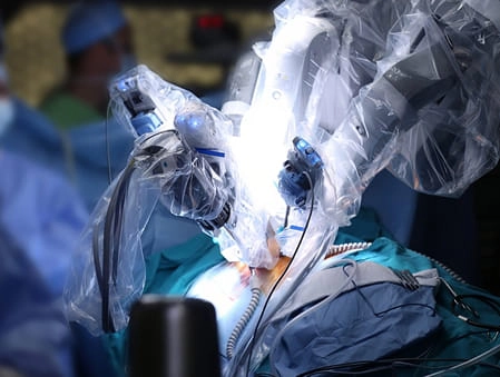
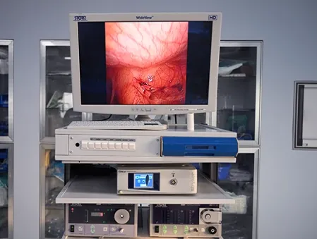
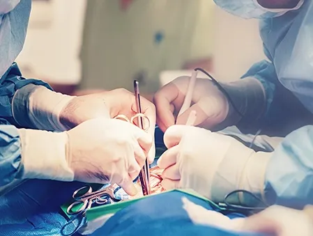
- Robotic Chest Surgery
- Video-assisted Thoracoscopic Surgery (VATS)
- Chest Wall Fixation for Chest Trauma
HITHOC (Hyperthermic Intrathoracic Chemotherapy) for Advanced Thymomas
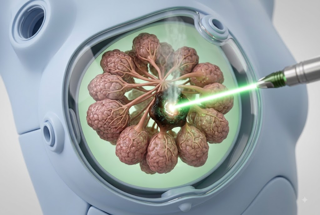
Hyperthermic Intrathoracic Chemotherapy (HITHOC) is a specialised intraoperative procedure used in the management of advanced thymomas, particularly those with pleural dissemination - a pattern of disease spread where tumour deposits seed the pleural surfaces and cannot be fully resected by conventional surgery alone. The technique involves surgically removing all visible tumour from the pleural cavity (cytoreductive surgery), then circulating a heated chemotherapy solution directly through the chest cavity for 60 to 90 minutes before closure. Heat potentiates the cytotoxic effect of cisplatin against residual microscopic disease, while the direct intracavitary delivery achieves drug concentrations far higher than intravenous administration could safely sustain. Medanta is one of a very small number of centres in India to offer HITHOC as a standard surgical option, and it is integrated into a comprehensive thymoma treatment pathway that includes preoperative staging, multidisciplinary tumour board review, and long-term oncological follow-up.
Patient Stories
It showcases inspiring journeys of patients who have received exceptional care at Medanta.


Diaphragmatic Eventration Treatment
Mr. Swastik

Abscess (Empyema)
Dr. Poonam Singla

Esophageal Fistula Treatment
Ms. Mansi Khupchandani
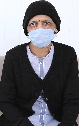
Esophageal cancer
Mrs. Anshika's Mother

Mucormycosis ( Black Fungus)
Mrs. Sarah Mehra