What Is Echocardiography and How Does It Work?
Published on: Nov 25, 2022
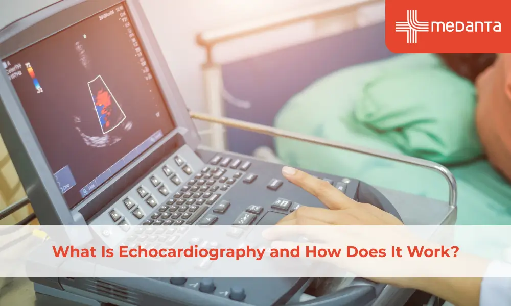
TABLE OF CONTENTS
Echocardiography uses a handheld probe to emit ultrasonic waves in your body. When the sound waves get reflected off the surface of your heart and are captured back with the probe, this helps create a visual representation of the graphic outline of your heart and its movements. An echocardiogram will be performed by a doctor called a cardiac sonographer. These doctors are trained specifically in performing echocardiography with the best technology and know how to produce the best results accurately.
There are several particular types of echocardiograms based on the technology used. These includes:
Types of Echocardiograms Based on Technology
M-mode echocardiography: This produces an image similar to a trace of the heart's borders. It is the simplest form showing the heart chambers, the size of the heart, and the thickness of the heart walls.
Doppler echocardiography: This procedure is specifically used to understand blood flow through the heart’s chambers and valves. It helps the doctor get a clearer picture of the heart's functioning.
Colour Doppler: Color Doppler takes Doppler echocardiography, goes one step further and uses different colours to indicate different directions of flow of blood through the heart.
Two-dimensional echocardiography: This technique helps to see the actual motion of rigid structures in real-time.
3D echocardiography: This method captures three-dimensional views of the heart with greater detail than two-dimensional echocardiography. 3D echocardiography is often used to plan the course of treatment for a heart condition.
How Does an Echocardiogram Help Diagnose Heart Problems?
Your doctor may order an echocardiogram for different reasons. For example, if you have an irregular heartbeat or your doctor suspects your heart is not pumping as it should, they try to detect the reasons using an echocardiogram. Here are some ways that doctors can understand the functioning of the heart better using an echocardiogram:
It helps the doctor understand changes in the size of your heart, its chambers, and the dilation and thickening of its walls.
It helps to detect whether their blood clots in the heart chambers.
It helps to detect fluid in the pericardial sac.
It helps to detect problems with the aorta, the significant blood vessel that carries blood from your heart.
It helps to understand the functioning of the heart and its contraction and relaxation phases.
It helps to understand problems with heart valves.
It helps to understand problems with pressure in the heart.
It helps to understand the health of the heart muscle, especially after a heart attack.
It can also detect congenital disabilities or inborn defects.
What are the conditions for which echocardiography may be suggested?
Cardiomyopathy – Ischemic or non-ischemic
Pericardial Effusion And Pericarditis
Atrial & Ventricle Septal Wall Defect
Heart Failure
Aortic Aneurysms & Aortic Dissection
Congenital Heart Disease – Cyanotic or Acyanotic
Problems With Heart Valves
Different Types of Echocardiography Procedures
There are several types of echocardiography based on the procedure.
Transthoracic echocardiography
This type of echocardiography uses a probe or a transducer that is placed over the chest. This transducer sends ultrasound waves toward your heart. The transducer captures the sound waves that bounce off the surface of your heart. A computer converts this information into visual images, which show the pumping of a heart as live images on the monitor.
Transesophageal echocardiography
This procedure uses a much smaller transducer, which is introduced down your throat through your mouth. Before this process, your doctor will numb the throat by a local anesthetic spray to make it easier and eliminate discomfort. This tube is passed through the oesophagus or the food pipe towards the back side of your heart. This transducer helps with a better view and visualise problems with some heart chambers better.
In this process, your doctor may give you a sedative to help you to relax before starting. Your doctor will also be giving an anaesthetic gel spray on your throat. The tube is then gently inserted, taking care not to create injury and ensuring that you will not feel any pain during the procedure or difficulty breathing. The process takes around half an hour.
Stress echocardiography
This procedure is similar to transthoracic echocardiography, but the doctor will take images before and after you have been asked to exercise or given medication eg- Dobutamine. This helps your doctor understand how the functioning of your heart changes according to stress.
3D echocardiography
This procedure uses a probe over your chest or oesophagus to create a 3D image of your heart. The computer combines multiple images from different angles to create a 3D image. It is also used to detect problems in the heart which are present at birth.
Fetal echocardiography
Fetal echocardiography is used when the pregnancy is 8 to 22 weeks in progress. For fetal echocardiography, the transducer probe is placed over the abdomen of a pregnant person. This test is safe because it does not use harmful radiation like X-rays.
Are There Any Risks with Echocardiography?
If your procedure involves using contrast dye, it may cause complications like allergic reactions in some people. Therefore, contrast is not used in pregnancy.
In some people, the trans-oesophageal probe causes a slight irritation in the throat. This may lead to a mild sore throat.
A stress echocardiogram temporally elevates the heart rate and BP to uncomfortable levels or causes irregular heartbeat in people with heart problems. This procedure is usually done with supervision to mitigate all risks.


