Femur Bone: Anatomy, Structure, Functions, Fracture Types, Treatment & Recovery
Published on: Apr 27, 2026
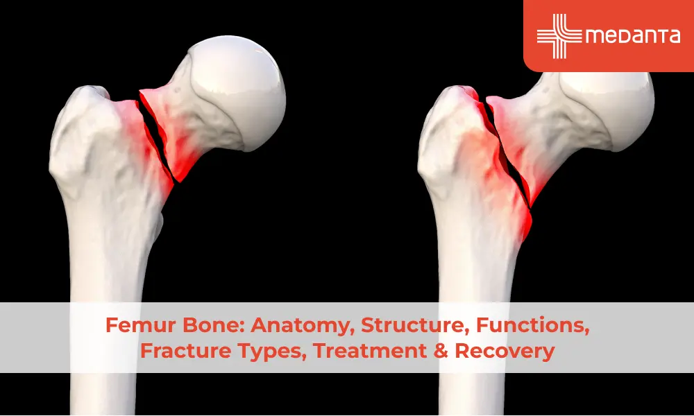
TABLE OF CONTENTS
- What Is the Femur Bone?
- Femur Bone Anatomy
- Femur Bone Structure
- Functions of the Femur Bone
- Common Femur Bone Problems & Pain Causes
- Femur Bone Fracture: Types & Symptoms
- Femur Bone Fracture Treatment Options
- Femur Bone Fracture Recovery Time
- Femur Bone Surgery & Bone Grafting
- When to See an Orthopaedic Specialist
- Conclusion
- FAQs
No bone in the body takes on more load than the femur. It carries the entire weight of the upper body, absorbs impact with every step and anchors most of the major muscles that move the hip and knee. It is also the most commonly fractured large bone in trauma and one of the more serious injuries to manage when it does break.
What Is the Femur Bone?
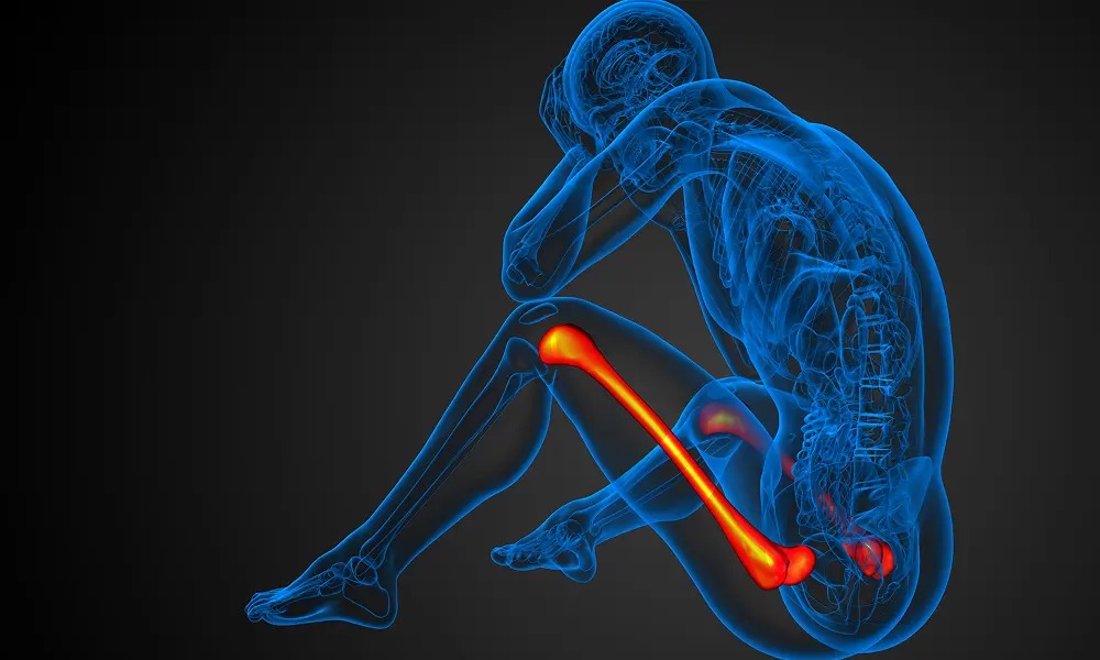
The femur is the single bone of the thigh. It connects the hip joint above (where the femoral head sits in the acetabulum of the pelvis) to the knee joint below, where the condyles articulate with the tibia. In a healthy adult male it averages around 48 cm in length. It is the longest bone in the body and also the strongest.
Femur Bone Anatomy
Working from top to bottom: the femoral head sits inside the hip socket. Below it is the femoral neck, a narrow bridge that is the most common fracture site in older adults. The greater and lesser trochanters are the bony projections where the gluteal and iliopsoas muscles attach. The shaft is the long cylindrical body. At the lower end, the medial and lateral condyles meet the tibial plateau to form the knee, with the patella sitting in the groove between them.
Femur Bone Structure
Two tissue types, layered by function. The outer shell (cortical bone) is dense and resists bending. The interior (trabecular or cancellous bone) is a lattice structure that handles compressive load with less weight. Running through the shaft is the medullary canal, which holds bone marrow. The periosteum, a fibrous membrane on the outer surface, contains the cells that initiate fracture repair. This is the layer surgeons protect during fixation.
Functions of the Femur Bone
Weight-bearing and locomotion are the obvious functions. Less obvious: the femur shields the femoral artery, the main blood supply to the lower limb, which runs along the posterior aspect of the shaft. A femoral shaft fracture can lacerate this vessel. That is one reason femoral shaft fractures are treated as surgical emergencies. The marrow also contributes to systemic function, red blood cell production continues in the femur throughout adult life.
Common Femur Bone Problems & Pain Causes
Fracture dominates the clinical picture for obvious reasons. Stress fractures develop from repetitive loading in runners and military recruits when microdamage accumulates faster than the bone can repair it. The femoral neck is the most common site and can complete if missed.
Avascular necrosis of the femoral head is a different problem as disruption of blood supply causes bone tissue to die and the femoral head eventually collapses. It follows femoral neck fractures, prolonged corticosteroid use and chronic alcohol use. Night pain without trauma, particularly in a patient on long-term steroids, should raise this possibility. Bone tumours (primary and metastatic) also present similarly.
Femur Bone Fracture: Types & Symptoms
Fracture Type | Location | Common Cause |
Femoral neck | Proximal, below the head | Falls, osteoporosis |
Intertrochanteric | Between trochanters | Low-energy falls, elderly |
Subtrochanteric | Below the lesser trochanter | High-energy trauma, pathological |
Shaft fracture | Mid-diaphysis | Road accidents |
Distal femur | Above knee, condyles | Direct trauma, sports |
Stress fracture | Shaft or neck | Repetitive loading, athletes |
Shaft fractures can cause up to 1.5 litres of internal bleeding into the thigh. Haemodynamic status needs assessing immediately alongside the orthopaedic injury.
Femur Bone Fracture Treatment Options
Surgery is the standard for adults. Traction and bed rest are largely historical.
Intramedullary nailing: A rod passed through the medullary canal and locked at both ends. Standard for shaft fractures. Allows early weight-bearing; union rates are excellent.
Dynamic hip screw: A sliding screw-plate for intertrochanteric fractures. The sliding component allows controlled compression as the patient loads the limb.
Hip replacement: For displaced femoral neck fractures in elderly patients where fixation failure risk is high. Replacing the joint is more reliable than trying to repair it in this group.
Plate fixation: For distal femur fractures and complex proximal patterns where nailing is not appropriate.
Femur Bone Fracture Recovery Time
Fracture Type | Union Time | Full Recovery |
Femoral neck (fixation) | 3–6 months | 6–12 months |
Femoral neck (replacement) | N/A | 3–6 months |
Intertrochanteric | 3–4 months | 4–6 months |
Shaft fracture | 4–6 months | 6–12 months |
Distal femur | 3–4 months | 6–9 months |
Femur Bone Surgery & Bone Grafting
Bone grafting is used when a fracture gap cannot bridge naturally due to comminution, bone loss or established non-union. Autograft from the patient's iliac crest is the gold standard; the patient's own bone provides the best biological environment for healing. Allograft from a bone bank, synthetic substitutes and demineralised bone matrix are used when autograft volume is insufficient. The graft is a scaffold. The patient's own osteoblasts colonise it and replace it with new bone over months.
When to See an Orthopaedic Specialist
Contact a doctor if you experience:
Severe thigh pain and inability to bear weight after trauma
Deep groin or thigh pain in a patient with osteoporosis, cancer history or long-term steroid use without obvious trauma
Night pain in the thigh unexplained by activity
An athlete whose thigh pain does not settle after two weeks of rest
Any new leg length difference or visible deformity.
Conclusion
The femur is not just the longest bone, it is the one the body depends on most for movement and load transfer. When it fractures, the consequences are serious and the management demands surgical expertise, careful rehabilitation and realistic timelines. Modern implants have transformed what is possible after femur fractures, but recovery is still measured in months, not weeks. If you are dealing with a femur fracture or unexplained thigh pain, a specialist assessment is the starting point.
FAQs
What is the femur bone and where is it located?
The single bone of the thigh, running from the hip joint above to the knee joint below. The longest and strongest bone in the body.
What are the main functions of the femur bone?
Weight-bearing, locomotion through muscle attachment, protection of the femoral artery, and red blood cell production in the marrow throughout adult life.
How strong is the femur bone compared to other bones?
It withstands forces up to 1700 newtons under normal loading and requires significant force to fracture in a healthy adult. In osteoporotic bone that threshold drops substantially, which is why elderly patients fracture with low-energy falls.
What are the common types of femur bone fractures?
Fracture types are:
Femoral neck fracture
Intertrochanteric fracture
Subtrochanteric fracture
Shaft fracture
Distal femur fracture.
Each has a distinct typical cause, surgical approach and recovery timeline.
How long does femur bone fracture recovery take?
Three to twelve months depending on fracture type and patient factors. Bone union comes first; full functional recovery with physiotherapy takes longer.
What is the treatment for a femur fracture?
Surgical in most adults. Intramedullary nailing for shaft fractures, dynamic hip screw for intertrochanteric, hip replacement for displaced neck fractures in the elderly and plate fixation for distal fractures.
When is femur bone surgery needed?
For almost all displaced femur fractures in adults. Early surgery reduces complications from immobility and produces better outcomes than prolonged conservative management.
What is bone grafting in femur fractures?
Filling a bone defect that cannot heal on its own due to comminution, bone loss or non-union. Autograft from the patient's own pelvis is most effective; donor bone and synthetic substitutes are used when autograft is insufficient.
Why does femur bone pain occur?
Femoral bone pain can occur due to fractures, stress fractures, avascular necrosis, hip or knee osteoarthritis, bone tumours, or referred lumbar spine pain. The pattern of onset, location and associated symptoms distinguishes between these.
What is the difference between femur bone anatomy and femur bone structure?
Anatomy is the named parts and their spatial relationships like head, neck, trochanters, shaft, and condyles. Structure is the tissue composition like cortical bone, trabecular bone, medullary canal, periosteum, that determines mechanical behaviour.