Ultrasound Scan: How It Works, What It Detects & Why It’s Safe
Published on: Jul 31, 2025
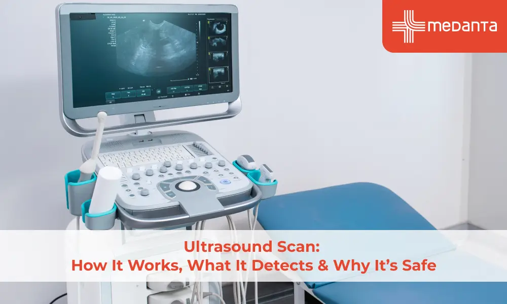
TABLE OF CONTENTS
Sound waves above 20 kilohertz create images of the body's internal structures through ultrasound technology. Doctors worldwide rely on this remarkable imaging technique, especially when they need to know about internal organs or monitor baby development during pregnancy.
Ultrasound's ability to capture immediate images sets it apart from other medical tools. Medical staff can observe actual organ movement and blood flow through vessels in detail. The technology's excellent safety record and absence of ionising radiation make it perfect for examining soft tissues that x-rays struggle to reveal clearly. The versatile technology helps diagnose many conditions through a process most patients find comfortable and non-invasive.
This article covers essential information about ultrasound examinations. You'll learn how they work, what happens during the procedure, available types, and the medical conditions they can detect.
What is an Ultrasound?
Sonography, also called ultrasound, creates images of what's inside the body using high-frequency sound waves. Medical ultrasounds come in two types: diagnostic and therapeutic.
Diagnostic ultrasound shows pictures of internal structures without any invasive procedures. This gentle technique lets doctors see organs, tissues, and blood flow through up-to-the-minute data analysis. So doctors can learn about how the body functions.
Therapeutic ultrasound doesn't make images but changes or destroys tissues instead. This method helps move tissues, dissolve blood clots, deliver drugs, or remove tumours without surgical cuts.
How It Works
A handheld device called a transducer contains piezoelectric crystals. These amazing materials turn electrical energy into sound waves and back again.
The sonography process looks like this:
Puts special gel on the skin to stop air pockets from forming between the skin and the transducer
Glides the transducer over the area being checked
The transducer sends sound waves into the body at 1-20 MHz frequencies
These waves bounce back as echoes after hitting tissues
The transducer catches these returning echoes and turns them into electrical signals
A computer turns these signals into images you can see on screen
The echo's strength changes based on how dense the tissue is. That's why bones look bright white and fluid appears black.
About Test
Doctors recommend ultrasound tests to break down symptoms, track existing conditions, or guide medical procedures. The scan usually takes 10 to 45 minutes, though some cases might need extra time.
A sonographer performs these scans with their specialised training. The procedure follows these steps:
They help you lie down on an examination table
They apply a special gel to the scan area
They slide a transducer on your skin to get images
They watch the results on a screen nearby
Some examinations need internal ultrasound probes. These include transvaginal ultrasounds (vagina), transrectal ultrasounds (rectum), and transesophageal echocardiograms (oesophagus).
Your scan's preparation depends on the body part being checked. Some tests need no preparation at all, while others might ask you to:
Skip eating for several hours
Fill your bladder with water
Hold your urine until the test ends
Ultrasound helps find many health issues. It can spot the causes of pain, detect blockages in blood vessels, tell the difference between cysts and solid tumours, and check blood flow. This versatile tool also helps check organs of all types like the heart, liver, kidneys, thyroid, breast and reproductive organs.
Your results might be ready right away or take a few weeks, based on your case.
What Conditions Can be Detected by Ultrasound?
Ultrasound's diagnostic capabilities work across body systems and serve as a great tool in modern medicine. This quickest way of imaging helps doctors tell the difference between fluid-filled cysts and solid tumours. Doctors can detect potential cancers early with this technology.
Ultrasound shows remarkable results for imaging palpable abnormalities during breast examinations. Dense breast tissue makes this technique particularly valuable since mammography's sensitivity drops substantially.
Ultrasound plays a vital role for expectant mothers:
Confirming pregnancy and checking for complications like ectopic pregnancy
Determining gestational age and due dates
Monitoring baby's growth, movement and heart rate
Detecting potential congenital conditions
Doctors use ultrasound to assess coronary heart disease by identifying abnormal myocardial activity. Echocardiograms show blood flow through heart valves and help diagnose conditions like valve disease and heart failure.
Doctors can detect many conditions through abdominal ultrasound, including kidney stones, liver disease, gallstones, pancreatitis and abdominal tumours. The technology also reveals abdominal aortic aneurysms and bladder stones.
Ultrasound proves its versatility by helping diagnose thyroid abnormalities and musculoskeletal injuries. It guides needle placement for biopsies, making it one of medicine's most versatile diagnostic tools.
Conclusion
Ultrasound technology is a remarkable medical breakthrough that lets doctors view our bodies' insides without surgery. This technique's safety is its biggest advantage - patients can get multiple scans without worrying about harmful radiation. Medical staff can watch organs function live instead of just looking at static images.
Most people think of pregnancy when they hear "ultrasound," but this versatile tool does so much more. Doctors use it to spot heart valve issues and locate kidney stones, making it a vital diagnostic method. The whole process is quick and comfortable for most patients and usually takes less than an hour.
Doctors value ultrasound's power to tell the difference between harmless fluid-filled cysts and potentially dangerous solid tumours. This distinction often leads to earlier treatment and improved outcomes. The technology helps doctors guide needles with precision during delicate procedures.
Ultrasound shows modern medicine at its most available form. Its combination of safety, speed, and diagnostic capabilities explains why doctors worldwide rely on this innovative technology. Your medical team can make better decisions about your care, whether you need a routine pregnancy check-up or answers about unexplained symptoms.
FAQs
What can an ultrasound detect?
Ultrasound helps identify many conditions, such as kidney stones, gallstones, liver problems, tumours, thyroid disorders, and heart valve issues. It also checks how a baby is growing during pregnancy.
Is ultrasound safe?
Yes, ultrasound is considered very safe. Unlike X-rays, it does not rely on ionising radiation. This makes it a good option to use often even while someone is pregnant.
How long does an ultrasound take?
Ultrasound exams take anywhere from 10 to 45 minutes. The time can change based on what part of the body is being checked or the kind of scan being done.