Carotid Cavernous Fistula (CCF): Causes, Symptoms & Treatment
Published on: Dec 05, 2025
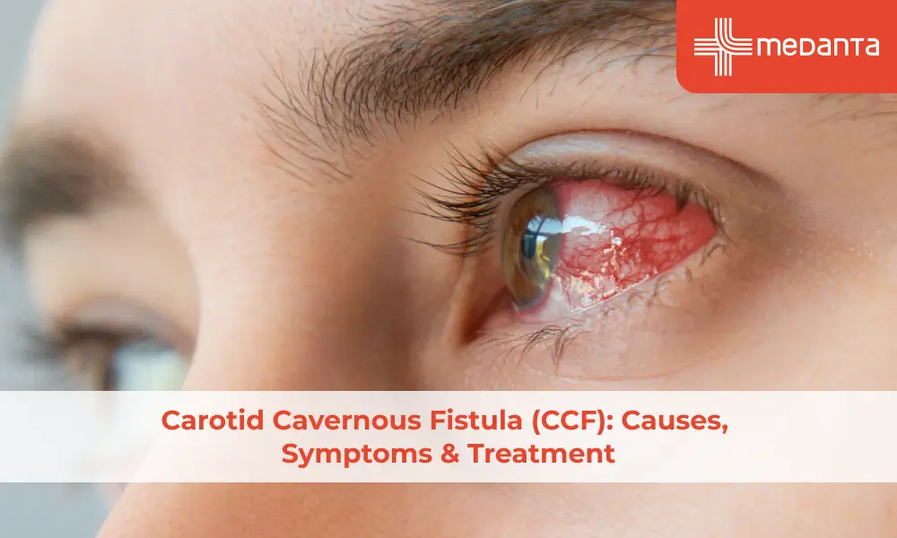
TABLE OF CONTENTS
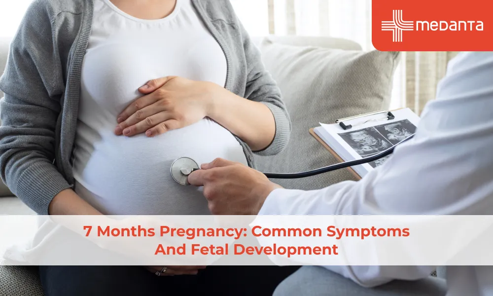
Carotid cavernous fistula (CCF) is a rare condition that needs immediate medical attention. CCF occurs when the carotid artery forms an abnormal connection with the cavernous sinus and disrupts blood flow patterns in the brain. Trauma causes most of these fistulas. Skull fractures and injuries lead to 70% to 75% of all cases. The remaining 30% are spontaneous CCFs that develop from aneurysm ruptures or genetic conditions. Patients show three classic symptoms: their eyes bulge out, they hear unusual sounds (whooshing sound) in their head, and the area around their eyes swells. This detailed guide looks at different types of this condition, including direct and indirect carotid cavernous fistulas.
What is Carotid Cavernous Fistula (CCF)?
A carotid cavernous fistula creates an unusual vascular connection between the carotid arterial system and the cavernous sinus. Blood flow patterns change when this abnormal shunting forces blood through alternative pathways in the skull.
Doctors have different ways to classify CCFs.
Direct fistulas connect the internal carotid artery to the cavernous sinus, which leads to high-flow, high-pressure conditions.
Indirect fistulas involve branches of either the internal or external carotid arteries.
Barrow's classification system, which is accessible to more people, divides CCFs into four types:
Type A: Direct connection between the intracavernous internal carotid artery and the cavernous sinus
Type B: Dural shunt between intracavernous ICA branches and cavernous sinus
Type C: Dural shunt between external carotid artery branches and cavernous sinus
Type D: Combination of types B and C
The cavernous sinus's location is vital because it contains essential neural structures, specifically cranial nerves III, IV, V1, V2, and VI. These nerves control eye movement and facial sensation, among other significant functions that the fistula might affect.
Bilateral CCFs are rare and appear in only 1% of traumatic cases.
Causes & Risk Factors
Causes
Carotid cavernous fistula causes are:
Traumatic injuries make up 70-75% of all cases. It includes:
Skull base fractures
Acceleration-deceleration forces
Penetrating head trauma
High-flow, direct fistulas commonly affect young males after such incidents.
About 30% of CCF cases happen spontaneously. Post-menopausal women tend to develop these low-flow, indirect connections more often. These spontaneous cases usually result from:
Rupture of an intracavernous carotid aneurysm
Genetic predispositions affecting arterial walls
Collagen disorders
Risk factors
CCF risk increases by a lot in people with certain genetic conditions, such as:
Ehlers-Danlos syndrome type IV
Fibromuscular dysplasia
Pseudoxanthoma elasticum
Osteogenesis imperfecta
Certain medical conditions increase the risk of CCF like:
Hypertension
Atherosclerotic disease
Pregnancy
Excessive straining
CCFs can also occur as complications from medical procedures, including:
Endovascular interventions
Trans-sphenoidal surgeries
Endoscopic sinus procedures
Carotid endarterectomy
Percutaneous treatment of trigeminal neuralgia
Symptoms of CCF
People with Carotid Cavernous Fistula show distinct symptoms that vary based on their condition's type and severity.
The classic "Dandy triad" consists of:
Sudden onset proptosis (bulging eyes)
Presence of a bruit (abnormal sound)
Conjunctival congestion (redness)
Direct CCFs emerge suddenly with all three elements of this triad. Indirect CCFs, however, progress slowly with symptoms that come and go, which makes diagnosis challenging.
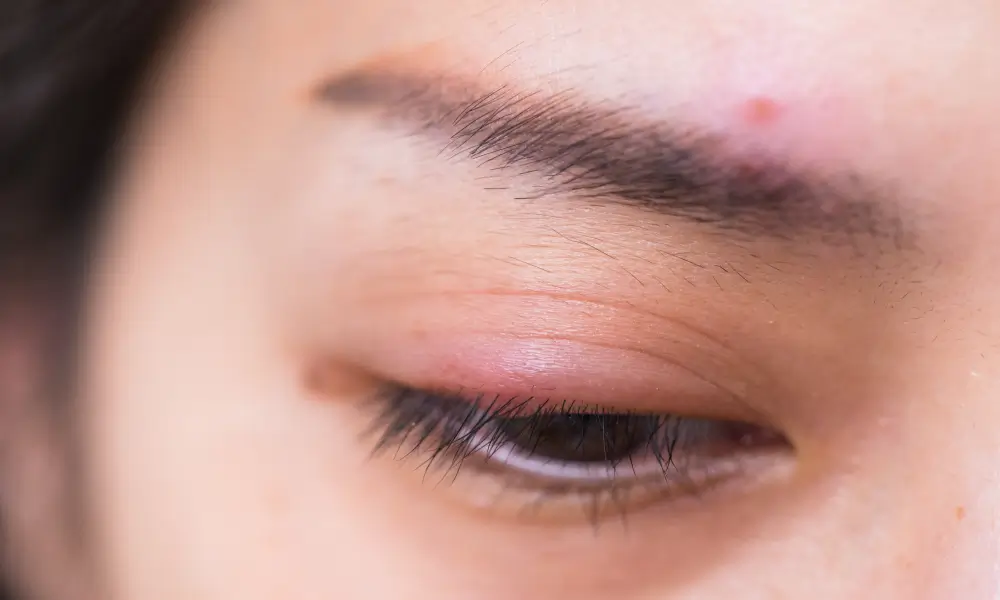
Eye-related symptoms stand out as the main clinical signs, and proptosis (bulging of the eyes) affects every patient. Other eye problems are:
Chemosis (swelling of eye membranes)
Engorged vessels
Diplopia (double vision)
Ophthalmoplegia (eye movement limitations)
Other symptoms of CCF are:
Headaches
Hearing problems like tinnitus
Eye-related sounds as "swishing" or "buzzing".
Vision problems range from slight blurring to severe loss. High pressure inside the eye disrupts aqueous humour drainage and causes these issues.
Uncommon symptoms include:
Nosebleeds
Hemiparesis
Brain bleeding
Diagnosis
Diagnosing a carotid cavernous fistula requires careful clinical assessment, along with advanced imaging.
Clinical examination: Doctors listen for ocular bruit during examination—a distinctive sound they detect with a stethoscope placed over the affected eye. This finding suggests the need for additional testing.
Diagnostic tests include:
Tonometry: Shows increased ocular pulse amplitude on the affected side
Ultrasound: Reveals dilated superior ophthalmic vein (SOV) and orbital congestion
CT and MRI scans: Display enlarged cavernous sinus, dilated SOV, and extraocular muscle enlargement
CTA and MRA: Detect CCFs causing visual symptoms with high sensitivity
Cerebral angiography: It is the gold standard diagnostic test. This definitive test reveals the fistula's exact location, feeding vessels and drainage patterns. Digital subtraction angiography (DSA) provides the clearest visualisation.
Treatment for Carotid Cavernous Fistula
Treatment for CCF depends on its flow speed and type. Treatment options are:
Endovascular interventions: These are the preferred treatment choice with remarkable success rates.
Transarterial embolisation achieves complete obliteration in the majority of cases for direct high-flow CCFs. This technique uses:
Detachable balloons
Platinum coils
Covered stents
Transvenous embolisation proves effective for indirect low-flow fistulas. Using coils alone in the transvenous approach yields higher success rates.
Both approaches deliver similar outcomes. Research shows no statistically significant differences between transarterial and transvenous techniques.
Non surgical approach: Some indirect CCFs on its own. These cases benefit from carotid self-compression done 4 times hourly to encourage thrombosis. Patients use their opposite hand to press the carotid artery for 20-30 seconds each time.
Surgical intervention: Doctors recommend surgery only after endovascular approaches fail. Surgical options are:
Direct suturing
Cavernous sinus packing
Carotid ligation
Radiosurgery serves as a valuable second-line option for indirect CCFs but requires months to show full results.
CCFs left untreated can lead to serious complications including visual loss, cranial nerve damage, and lasting cosmetic issues.
Prognosis and Recovery
Recovery outcomes after CCF treatment depend on multiple factors:
Recovery Aspect | Success/Recovery Rate | Timeline |
Complete occlusion (immediate) | High | Immediately after the procedure |
Complete remission (long-term) | High | Within 6+ months |
Ocular symptom improvement | High | Weeks to months |
Transvenous vs transarterial approach success | Higher vs moderate | Immediate obliteration |
Proptosis & chemosis resolution | Most cases | Hours to days |
Cranial nerve palsy recovery | Moderate | Several weeks |
Intraocular pressure normalisation | Significant improvement | Immediate to months |
Cranial nerve complications | Low | Varies |
Recurrence rates | Low | Variable |
Symptoms resolve in a predictable sequence. Patients experience immediate relief from ocular bruits and pulsations after successful treatment. Eye pressure returns to normal quickly, while conjunctival problems may take weeks or months to clear up completely.
The patient's visual recovery depends on three key factors: the fistula's flow rate, how quickly intervention happens, and whether the optic nerve has permanent damage.
Conclusion
Carotid Cavernous Fistula is a rare but vital vascular condition that just needs immediate medical attention. The telltale signs include bulging eyes, unusual head sounds, and swelling around the eye area.
Quick diagnosis is a vital step to prevent permanent vision loss. Doctors can determine the
best treatment by understanding direct and indirect fistulas. Treatment methods have evolved remarkably.
Recovery times differ based on the fistula type & treatment speed. With the right treatment most patients bounce back. CCF may be rare, but knowing its signs could mean the difference between complete healing and lasting damage. That's why patients should seek medical help at the first sign of symptoms.
FAQs
What causes carotid cavernous fistula?
Trauma usually causes direct CCFs through skull base fractures, acceleration-deceleration injuries, or penetrating head wounds. Indirect CCFs develop spontaneously when dural branches rupture due to hypertension, genetic conditions like Ehlers-Danlos syndrome, or collagen vascular disorders.
What are the early signs of CCF?
The original symptoms show up as redness in one eye, orbital pain, and buzzing sounds in the head. Direct CCFs cause sudden symptoms, while indirect fistulas progress slowly. Patients may experience eye bulging and double vision soon after.
How is CCF diagnosed?
Cerebral angiography remains the gold standard for diagnosis. Doctors can detect it earlier through tonometry that shows increased ocular pulse amplitude, B-scan ultrasound that reveals dilated superior ophthalmic veins, and CT/MRI scans that display orbital congestion.
Can carotid cavernous fistula go away on its own?
Yes - indirect CCFs close spontaneously in some cases. Direct high-flow fistulas rarely heal without treatment.
What is the success rate of CCF treatment?
Endovascular interventions succeed most of the time. Transvenous approaches achieve higher complete obliteration compared to transarterial methods.
What complications can arise from untreated CCF?
Untreated CCFs may cause:
Permanent visual loss
Persistent cranial nerve paralysis
Glaucomatous damage
Retinal vein occlusion
Intracranial haemorrhage
Is surgery always required for carotid cavernous fistula?
No. Endovascular procedures have become the preferred treatment option over traditional surgery. Low-flow fistulas respond to conservative carotid compression therapy about 30% of the time.
How long is recovery after CCF treatment?
Recovery time depends on your treatment type and personal factors. Most patients see quick improvements in ocular bruits and pulsations. Proptosis and chemosis get better within days. Cranial nerve palsies might take several weeks to clear up. Most patients see all their symptoms resolve within 6 months.
The fistula type plays a role in recovery speed. Direct CCFs tend to show faster improvement after successful embolisation than indirect cases.
Can CCF affect vision permanently?
If left untreated carotid cavernous fistula can damage your vision forever. Your risk goes up with:
Treatment delays (especially past 1 month)
High-flow direct fistulas
Pre-existing optic nerve compression
Secondary glaucoma development
Are there preventive measures for carotid cavernous fistula?
Head injuries cause most direct CCFs, so prevention focuses on reducing these risks. You should wear proper protective gear during risky activities and follow vehicle safety rules. People with Ehlers-Danlos syndrome should get regular vascular checkups to spot weaknesses early. Additionally, it helps to control blood pressure and avoid straining too hard, especially if you have a history of vascular issues.