THE EXCHANGE | Newsletter October 2018
Treating Ureteric Calculus in a Rare Anatomical Anomaly in a Child
A seven-year-old, otherwise healthy male, presented with right colicky flank pain for three days. He was voiding normally, with no history of urinary tract infection. His father had a positive history of being treated for renal calculi.
On investigation, renal function tests and metabolic work-up for stone were normal. Ultrasound showed mild right upstream hydroureteronephrosis with a rare finding of a
6 mm calculus near the ectopic vesico-ureteric junction in the prostatic urethra. The reported incidence of ureteral ectopia is 1 in 2000 children, although the actual incidence is difficult to determine.
A 6-week trial of alpha-blockers failed to expel the calculus, although the dilatation in the renal unit subsided.
A ureteroscopy was attempted elsewhere in a foreign country, but failed owing to non-visualization of the ectopic ureteric orifice (eUO).
After three months, with complains of recurrence of pain, the patient was brought to Medanta - The Medicity. A CT urography with 3D reconstruction confirmed a 1050 HU, 6 mm calculus near the ectopic opening of the right ureter into the prostatic urethra. A dynamic renal scan showed preserved renal function with slow, but non-obstructed drainage.
Endoscopic removal of the lower ureteric calculus to avoid the pain and scar of an open surgical approach was planned. Given the rarity of the condition i.e. a single system ectopic ureter (SSEU) with lower ureteric calculus in a male child, a team of pediatric urologist, endourologist and interventional radiologist worked together to holistically treat the condition. The team developed an operative protocol.
A combined ultrasound and fluoroscopy-guided antegrade access into lower calyx of the mildly prominent right renal unit was obtained by the Intervention Radiology team.
The narrowing at the ectopic vesico-ureteric junction was crossed using a 5 Fr multipurpose angiocatheter (MPA). In the same anesthetic sitting, the case was then taken over by Pediatric Urology and Endourology teams. A 0.35” terumo guidewire was universalized by placing it through the MPA into the bladder and per-urethral retrieval cystoscopically. Controlled dilatation of the eUO was done using a 5Fr balloon dilator. A 7.5 Fr semi-rigid ureteroscope was then guided into the eUO. The calculus was embedded in odematous bullae, nearly 1 cm proximal to the eUO and was dislodged with the tip of the ureteroscope. It was retrieved using a stone holding forceps and a 4Fr, 20cms DJ stent placed into the bladder.
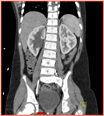
CECT (coronal section) showing calculus [red arrow] at the site of insertion of ectopic right ureter into the prostatic urethra.
The total operative time was 60 minutes. The patient was discharged on post-operative day-1, after removal of Foley's catheter and no resultant urinary leak. DJ stent was removed on the seventh day after surgery. On follow-up, the patient continues to void in a normal stream with no residual hydroureteronephrosis.
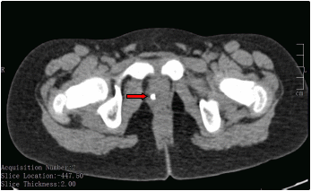
CECT (sagittal section) showing the calculus [red arrow] below the region of bladder neck, far from the opening of the normal ureteric orifice
Such collaborative work illustrates that endoscopic management of ureteric calculi in a male with SSEU is feasible and should be given careful consideration against the conventional open ureteric reimplantation. The open approach is limited by space constraints in a pediatric
pelvis and difficulty in approaching the sphincteric intra-mural portion of the ectopic ureter in males, culminating in high failure rates for stone retrieval, despite undertaking an extensive surgery. In contrast, the evolution of finer endoscopic instruments for pediatric urolithiasis now permit the benefits of minimally invasive surgery to be extended to a larger, formidable spectrum of pediatric patients.
Endourology can thus offer a one-stop, single stage successful intervention to the challenging diagnosis of lower ureteric calculus in an ectopic ureter. A coordinated effort between pediatric urology, endourology and intervention radiology teams, detailed work-up and meticulous planning are critical to treating such patients.
https://www.medanta.org/dr-virender-kaur-sekhon/
https://www.medanta.org/dr-manav-suryavanshi/
https://www.medanta.org/dr-abhay-kapoor/
Vein-to-Vein Safety of Blood Transfusion
At the Medanta Blood Bank, we have safeguards and quality assurance governing the entire process of blood donation-to-blood transfusion. The process normally involves collection of whole blood from a healthy donor, processing it into components, testing each donation for Transfusion Transmitted Infections (TTI), doing pre-transfusion compatibility testing and finally, doing bed-side checks before transfusing it into the patient.
Donor counseling
Blood Transfusion Services (BTS) team incorporates counseling before, during and after donation to ensure safety of blood donor and sustain a healthy pool of repeat donors. We recognize pre-donation counseling as one of the elements to prevent or reduce the donation of blood by donors who might be at risk for TTI, as well as to give them an understanding of the entire donation process. Counseling during and immediately after donation also contributes to donor safety by reducing the chances of adverse reaction(s).
Leukodepletion
We also obtain an informed consent from the donors that their blood samples will be tested for TTI like Human Immunodeficiency Virus (HIV), Hepatitis B Virus (HBV), Hepatitis C Virus (HCV), Syphilis and Malaria, and to ascertain whether they wish to be informed about a positive test result. Post-donation counseling is done to inform donors of unusual or abnormal test results.
Donation process
We have a stringent medical history questionnaire and brief physical exam to screen out (defer) donors who may not be fit for donation. Governing principle is that “neither donor, nor recipient should be harmed”. At the time of blood collection we use special bags with pre-sampling pouch to collect initial few milliliters of blood for ensuring safety of the collected blood. Very few centers in India follow this practice.
Optimal utilization of donated blood
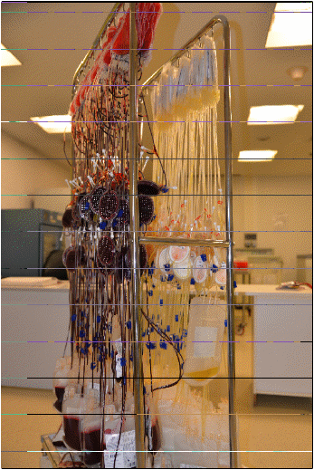
Each unit of whole blood is split into components; Red Blood Cells (RBC), Platelet Concentrate (PC) and Fresh Frozen Plasma (FFP); which is the scientific way of using blood clinically and ensuring that the scarce resource donated by
a donor is used optimally to benefit multiple patients. Moreover, each RBC and PC is leucodepleted (removal of White Blood Cells [WBC]) to enhance patient safety by reducing the adverse reactions caused by WBC. In India, 100% leukodepletion of all RBC and PC units is done only at Medanta.
Rigorous testing
In line with the global best practice, each unit undergoes two-tier testing. In the first tier, Chemiluminescence technology is employed for testing anti-HIV, anti-HCV, antibodies to Syphilis and HBsAg. Immuno-chromatography is used for testing Malaria.
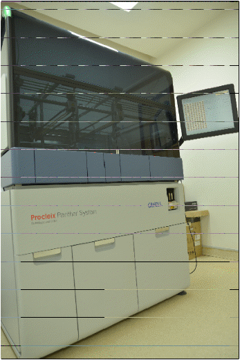
The second tier comprises of Nucleic-acid Amplification Test (NAT) testing for individual units by an automated processor called Panther. NAT testing is only practiced in about 10% of all blood-banks in the country.
Pre-transfusion compatibility testing
Medanta's blood bank is one of the handful in India that
does antibody screening in all admitted patients besides
the usual blood ABO and RhD grouping. This offers the advantage of augmenting patient safety by detecting presence or absence of irregular antibody in a patient.
If an antibody is detected, it is identified and corresponding antigen-negative blood unit is reserved for the patient.
The blood bank at Medanta is possibly the only one in the country which does only Immediate Spin Test before final issue of RBC to patients.
https://www.medanta.org/dr-aseem-kumar-tiwari/
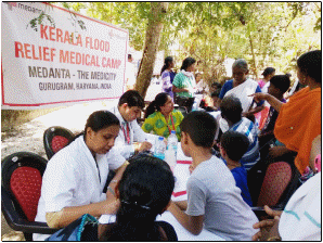
Medical Relief to Flood-Hit Kerala
A medical team comprising of eleven volunteers from Medanta - The Medicity travelled to Kerala to provide medical aid and support to flood-hit areas.
Over a period of eleven days, the team provided medical services and distributed medicines to people suffering the aftermath of the devastation. Camps were held in Kochi and Chenganoor, the worst affected by the flood. In these camps, nursing care was provided, home visits were done for immobile old people, counseling sessions were held and mosquito repellents and sanitation material was distributed. Over 3000 people were served by our doctors and nurses.