1068
1068
 Request Call Back
 Contact
 1068
1068
Endotracheal Tube (ET Tube): Parts, Types, Uses and Insertion Procedure
Published on: Sep 24, 2026
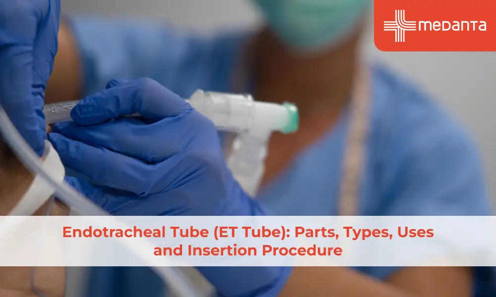
TABLE OF CONTENTS
An endotracheal tube (ET tube or ETT) is a flexible plastic tube inserted through the mouth or nose, passes through your vocal cords and into the trachea (windpipe) - all this is done to establish a secure airway. It is one of the most fundamental pieces of equipment in anaesthesia, critical care medicine and emergency medicine and is used whenever a patient's natural airway is compromised, inadequate or needs to be protected from aspiration during surgery or intensive care.
What Is an Endotracheal Tube?
An endotracheal tube is a hollow, cylindrical tube. It is made of polyvinyl chloride (PVC) which your doctor inserts into your trachea to create an airway. This pathway is a patent, protected pathway directly into the lungs that bypasses the upper airway. Once in position, the ET tube maintains an open airway in patients who cannot do so themselves, allows delivery of oxygen and anaesthetic gases, and protects the lungs from aspiration by inflating a balloon-like cuff that seals the trachea around the tube.
ET tubes are sized by their internal diameter in millimetres (ID). The typical sizes in clinical practice in India are 7.0 to 8.0 mm ID for adult men, 6.5 to 7.5 mm ID for adult women and 2.5 to 5.5 mm ID for paediatric patients.
Parts of an Endotracheal Tube
A standard cuffed endotracheal tube has several distinct components, each serving a specific function. They are
The tube body: The main transparent PVC shaft bearing centimetre markings along its length to confirm depth of insertion, typically 21 to 23 cm at the teeth in adult women and 22 to 24 cm in adult men
The bevel tip and Murphy's eye: The angled distal opening that facilitates smooth passage through the vocal cords, with a small lateral opening (Murphy's eye) near the tip that provides an alternative gas passage if the primary opening is occluded
The cuff: A balloon-like structure just proximal to the tip of the tube, inflated with air after insertion. When inflated, the cuff creates a seal that prevents aspiration and prevents gas from leaking around the tube during positive pressure ventilation
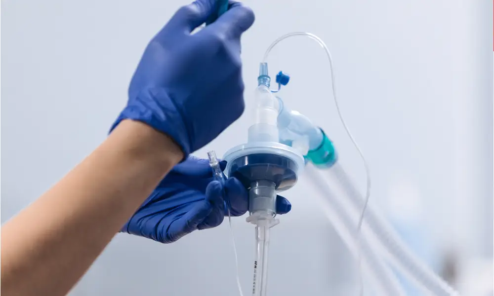
The pilot balloon and 15mm adapter: The pilot balloon is the small external balloon that allows assessment of cuff inflation state by palpation. The proximal end terminates in a standard 15mm universal connector that attaches to any compatible breathing circuit, ventilator or Ambu bag
The radio-opaque line: A thin line of radio-opaque material embedded in the tube wall that is visible on a chest X-ray and used to confirm ET tube tip position.
Types of Endotracheal Tubes
Several specialised types of ET tubes exist for specific clinical situations.
Standard cuffed PVC ET tube: The most widely used type and is suitable for most adult elective and emergency intubations.
Uncuffed ET tube: Used in paediatric patients below approximately 8 years of age. The narrow paediatric subglottic airway provides an adequate seal around an appropriately sized uncuffed tube, reducing the risk of cuff pressure injury.
Reinforced (armoured) ET tube: Contains a spiral-embedded wall and is used in neurosurgery, ENT surgery and prone positioning.
Preformed (RAE) tube: A pre-curved ET tube that routes the breathing circuit away from the surgical field - nasal RAE tubes are used in oral surgery, and oral RAE tubes are used in procedures involving the face or head.
Double-lumen ET tube: Used in thoracic surgery to allow independent ventilation of the left and right lungs.
Uses of Endotracheal Tube in Medicine
Endotracheal intubation is indicated in any clinical situation where the patient's airway cannot be maintained safely by other means or where controlled mechanical ventilation is required. The indications are
General anaesthesia: Maintains a safe airway during surgery and protects against aspiration
Respiratory failure requiring mechanical ventilation: For patients with severe pneumonia, ARDS, exacerbations of COPD, or pulmonary oedema that cannot be managed with non-invasive ventilation
Emergency airway management: In patients with reduced consciousness (GCS below 8), cardiac arrest, severe trauma with airway compromise, and anaphylaxis with impending airway obstruction
Endotracheal Intubation Procedure - Step by Step
Endotracheal intubation is a skilled procedure performed by trained doctors. The following are steps for oral intubation using direct laryngoscopy:
Pre-oxygenation: The patient is pre-oxygenated for three to five minutes with 100% oxygen via a tight-fitting face mask, providing a safe apnoea time during the intubation procedure.
Induction of anaesthesia or sedation: In elective procedures, the patient is anaesthetised with an intravenous induction agent. In emergency RSI a rapidly acting induction agent is given simultaneously with a rapidly acting neuromuscular blocking agent.
Positioning and laryngoscopy: The patient is positioned in the 'sniffing position' (slight neck flexion and atlanto-occipital extension) to align the oral, pharyngeal and laryngeal axes. The laryngoscope blade is inserted and advanced to reveal the glottic opening.
Tube insertion: The ET tube is passed through the vocal cords under direct vision until the cuff is 1 to 2 cm below the vocal cords, at the appropriate depth as confirmed by the centimetre markings.
Cuff inflation and confirmation: The cuff is inflated to 20 to 30 cmH2O. Tube position is confirmed by end-tidal CO2 monitoring (the gold standard for confirming tracheal placement), bilateral auscultation and chest X-ray.
Risks and Complications of ET Tube Insertion
Complications can occur during insertion, in the immediate post-intubation period, or during prolonged intubation.
Oesophageal intubation is the most immediately dangerous complication. If not recognised and corrected immediately, it causes hypoxia and cardiac arrest.
Advancement of the ET tube too far results in the tip entering one main bronchus (usually the right), thereby ventilating only one lung.
Ventilator-associated pneumonia (VAP) caused by microaspiration of contaminated secretions past the cuff during prolonged mechanical ventilation.
Sore throat and hoarseness are reported in many patients in the post-operative period.
More serious complications include arytenoid dislocation and tracheal mucosal damage from cuff overinflation.
What Is ETT? Common Terminology Explained
ETT stands for endotracheal tube. In clinical practice, the terms ET tube, ETT, endotracheal tube, tracheal tube, and breathing tube are used interchangeably. Intubation is the process of inserting the ET tube. Extubation is removal of the ET tube when the patient can maintain their own airway. Reintubation is re-insertion of the ET tube if the patient fails extubation.
Conclusion
The endotracheal tube is one of the most important devices in modern medical practice - essential to the safe conduct of general anaesthesia, the management of acute respiratory failure, and the resuscitation of critically ill patients. Endotracheal intubation is a skill-dependent procedure that requires thorough training and systematic confirmation of tube position after insertion. When performed by trained doctors using appropriate monitoring it is a safe and effective intervention that saves lives in some of the most critical clinical situations encountered in medicine.
FAQs
What is an endotracheal tube used for?
An endotracheal tube is used to establish a secure, protected airway in patients who cannot maintain their own airway or who require controlled mechanical ventilation. The main uses are: general anaesthesia; acute respiratory failure requiring invasive mechanical ventilation; emergency airway management in unconscious patients, cardiac arrest and airway obstruction; and airway protection from aspiration in patients with reduced consciousness.
What does ET tube stand for?
ET tube stands for endotracheal tube - 'endo' means within, and 'tracheal' refers to the trachea (windpipe). The abbreviation ETT (endotracheal tube) is used interchangeably with ET tube in clinical practice. Related terms used in clinical settings include tracheal tube, breathing tube and airway tube.
What are the parts of an endotracheal tube?
The main parts of a standard cuffed endotracheal tube are: the tube body (transparent PVC shaft with centimetre markings); the bevel tip (the angled distal opening, with Murphy's eye as a lateral safety opening); the cuff (inflatable balloon that seals the trachea to prevent aspiration and gas leak); the pilot balloon (the external balloon for assessing cuff inflation state); the 15mm universal adapter at the proximal end (for connection to breathing circuits and ventilators); and the radio-opaque line (embedded in the tube wall for X-ray confirmation of tube position).
What are the different types of endotracheal tubes?
The main types of endotracheal tubes are: the standard cuffed PVC ET tube (used for most adult intubations); the uncuffed ET tube (used in infants and young children below approximately 8 years of age); the reinforced (armoured) ET tube (prevents kinking - used in neurosurgery, ENT surgery, and prone positioning); the preformed RAE tube (pre-curved for oral or nasal positioning - used in head and neck surgery); and the double-lumen ET tube (used in thoracic surgery for independent ventilation of each lung).
How is endotracheal intubation performed?
Endotracheal intubation is performed in the following steps:
Pre-oxygenation with 100% oxygen for three to five minutes
Induction of anaesthesia or RSI with a rapid induction agent and neuromuscular blocking agent
Positioning of the patient in the sniffing position
Laryngoscopy to visualise the glottic opening
Passage of the ET tube through the vocal cords under direct or video-assisted visualisation
Inflation of the cuff
Confirmation of tracheal position by end-tidal CO2 monitoring, auscultation and chest X-ray.
What is the purpose of an endotracheal tube cuff?
The cuff is an inflatable balloon that creates a seal between the outer surface of the ET tube and the tracheal wall preventing aspiration of secretions, gastric contents or blood into the lungs and stopping gas from leaking around the tube during positive pressure ventilation.
What is ETT in medical terms?
ETT in medical terms stands for endotracheal tube - the same device as an ET tube, a tracheal tube or a breathing tube.
What are the risks of endotracheal tube insertion?
Endotracheal tube insertion has few risks. They are:
Oesophageal intubation
Dental and soft tissue injury during laryngoscopy
Endobronchial intubation
Laryngeal and tracheal mucosal injury
Ventilator-associated pneumonia (VAP) in patients requiring prolonged mechanical ventilation.
How long can a patient stay on an endotracheal tube?
A patient can remain on an endotracheal tube for as long as mechanical ventilation is required. However prolonged intubation increases the risk of ventilator-associated pneumonia, tracheal mucosal injury from cuff pressure and tracheal stenosis. When mechanical ventilation is anticipated for more than approximately 7 to 14 days, elective tracheostomy is generally considered as an alternative as it is better tolerated by the patient and has a lower complication profile.
What size endotracheal tube is used for adults?
For adult patients, the standard sizes used in clinical practice in India are 7.0 to 8.0 mm internal diameter (ID) for adult men and 6.5 to 7.5 mm ID for adult women. These sizes allow adequate gas flow and suction catheter passage while fitting within the adult tracheal lumen.
Can an ET tube cause a sore throat?
Yes sore throat is one of the most common complications of endotracheal intubation. It is caused by mucosal irritation from the tube during insertion and by sustained pressure on the pharyngeal and laryngeal mucosa during intubation. The sore throat is typically mild to moderate in severity, lasts for one to three days after extubation, and resolves without specific treatment.
References
1. Hagberg CA, Artime CA. Airway management in the adult. In: Miller RD, ed. Miller's Anesthesia, 8th ed. Elsevier. 2015
2. Weingart SD, Levitan RM. Preoxygenation and prevention of desaturation during emergency airway management. Ann Emerg Med. 2012;59(3):165–75. https://doi.org/10.1016/j.annemergmed.2011.10.008
3. El-Boghdadly K, Bailey CR, Wiles MD. Postoperative sore throat: a systematic review. Anaesthesia. 2016;71(6):706–17. https://doi.org/10.1111/anae.13438
4. Kalanuria AA, Zai W, Mirski M. Ventilator-associated pneumonia in the ICU. Crit Care. 2014;18(2):208. https://doi.org/10.1186/cc13775
5. Myatra SN, Shah A, Kundra P, et al. All India Difficult Airway Association 2016 guidelines for the management of unanticipated difficult tracheal intubation in adults. Indian J Anaesth. 2016;60(12):885–98. https://doi.org/10.4103/0019-5049.195481