
Ultrasound, a prominent form of sonography, creates pictures of your body's interior using high-frequency sound waves without invasion. The technique proves safer than X-rays since it doesn't use radi.....
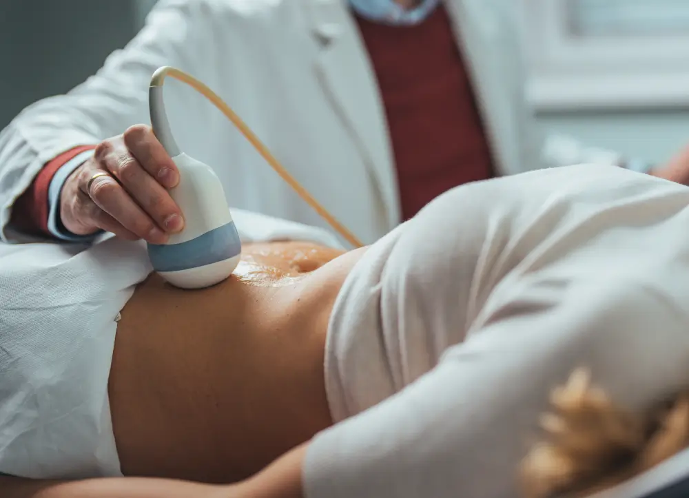
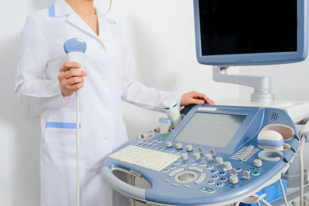
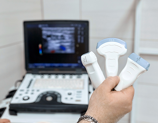
Ultrasound, a prominent form of sonography, creates pictures of your body's interior using high-frequency sound waves without invasion. The technique proves safer than X-rays since it doesn't use radiation.These remarkable sound waves work at frequencies above 20KHz, which human ears cannot detect. Medical applications typically use frequencies between 2 and 20 MHz.A doctor uses a handheld device called a transducer with a thin gel layer on your skin. The device's piezoelectric crystals transform electrical energy into sound waves. These waves move through your body at 1,540 metres per second, about one mile per second. The crystals switch to receive mode and catch echoes as waves return.
-
Ultrasound technology plays an important role in many fields and has become a powerful tool in today's world. Doctors use these sound waves to detect abnormalities without invasive procedures. Parents get their first look at their baby through an ult...
-
Doctors use ultrasound to get into internal organs like the heart, liver, kidneys and gallbladder. This technology effectively spots issues like tumours, cysts and inflammation. More importantly, cardiologists rely on echocardiograms to check heart f...
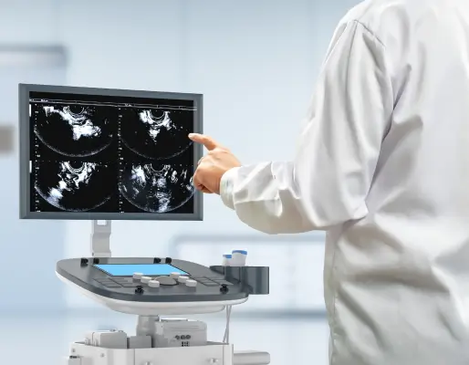
What Makes an Ultrasound So Unique?

Different body parts need different types of preparation before an ultrasound:
- Abdominal scans require 8-12 hours of fasting
- Clear images of the pelvis need you to drink one litre of water an hour before the scan
- Many exams need no preparation
Your doctor will tell you specific instructions about food, drinks, and medications before your appointment.
You'll change into a hospital gown when you arrive. The sonographer will ask you to lie down on an examination table and start the scan:
- A special water-based gel goes on your skin to eliminate friction
- The sonographer uses a device called a transducer against your skin
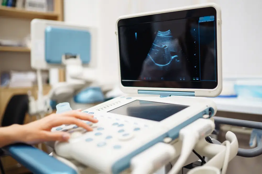
- Sound waves from the transducer travel through your body and create echoes
- Most scans take 20-40 minutes
Some exams might need an internal ultrasound. This method uses a special probe inserted through a natural body opening.

The sonographer will:
- Clean off the gel with paper towels
- Let you get back into your clothes
You can return to your normal routine right after the ultrasound. Your doctor will analyse the results with you during your next visit.
Ultrasound proves exceptionally safe because it doesn't use ionising radiation, which makes it suitable for pregnant women and children. This non-invasive approach needs no needles, injections, or incisions.
-
The technology gives users several advantages: Doctors can observe dynamic processes with immediate images The cost stays lower than other imaging methods like CT scans or MRI Soft tissues show up clearly compared to X-rays Patients need no recovery time after the procedure The process guides minimally invasive procedures perfectly The widespread availability helps bridge healthcare gaps in developing regions where expensive imaging equipment remains scarce.
-
The excellent safety record doesn't mean ultrasound comes without concerns. Potential risks include: Sound waves might heat tissues slightly or create small gas pockets (cavitation) Bone or air-filled structures block effective imaging Image quality depends on the operator's skill and experience Doctors should minimise exposure time and watch output settings carefully to ensure safe results.
Ultrasound's power comes from how easily it adapts to different needs. Unlike other imaging methods, this technology shows movement live, letting doctors see blood flow through vessels and watch organs work. This makes it a valuable tool for diagnosis and monitoring patients.
The technology is available to more people than other options. Many places that can't afford expensive CT and MRI machines use ultrasound to fill crucial healthcare gaps. Doctors can bring it right to the patient's bedside instead of moving them to special imaging rooms.
Key highlights are:
- It works without harmful radiation, which means doctors can use it more often
- Sound waves move through human tissue at 1,540 metres per second
- Most scans take just 30-40 minutes
- The device sends and receives sound waves, and spends 99% of its time "listening"
Ultrasound does have its limits. It can't see through bone or air-filled spaces well. That's why doctors need different imaging tools to look at the lungs or the brain.
Ultrasound combines safety, ease of use, and live imaging in a way that other technologies can't match.
