Placenta Previa: Symptoms, Causes, Diagnosis & Treatment
TABLE OF CONTENTS
- What is Placenta Previa?
- Types of Placenta Previa
- Symptoms of Placenta Previa During Pregnancy
- Causes and Risk Factors of Placenta Previa
- How Placenta Previa is Diagnosed
- Treatment Options for Placenta Previa
- Risks & Complications for Mother and Baby
- Delivery Planning in Placenta Previa
- Can Placenta Previa Resolve on Its Own?
- Precautions & Lifestyle Tips During Pregnancy
- FAQs
Placenta previa is one of the more significant obstetric complications of the second and third trimester. The diagnosis understandably unsettles pregnant women and their families. This article covers what placenta previa is, how it presents, how it is diagnosed and managed, and what delivery planning looks like when it is present.
What is Placenta Previa?
The placenta normally implants in the upper uterine segment, well clear of the cervical os. In placenta previa, the placenta implants abnormally low, partially or completely covering the internal os. This matters because the cervix must dilate for delivery; a placenta positioned over it creates a direct obstruction and a source of haemorrhage as the lower segment stretches and the cervix effaces. It complicates approximately 0.3–0.5% of pregnancies at term. Identified early, the position may change as the uterus grows; identified late, it requires specialist management and planned delivery.
Types of Placenta Previa
Complete (total) previa: The placenta covers the internal os entirely. Vaginal delivery is not possible. Caesarean section is mandatory.
Partial previa: The placenta covers part of the internal os. Degree of overlap determines obstetric management; Caesarean is usually required.
Marginal previa: The placental edge reaches the margin of the internal os but does not cross it. Depending on the distance (typically assessed on transvaginal ultrasound) vaginal delivery may be considered in carefully selected cases.
Low-lying placenta: The placental edge is within 2 cm of the os but does not reach it. This carries lower risk than true previa but still warrants monitoring and planned delivery at a centre with appropriate neonatal and surgical backup.
Symptoms of Placenta Previa During Pregnancy
The hallmark symptom is painless vaginal bleeding in the second or third trimester. Other symptoms are:
Sudden, bright red vaginal bleeding often without preceding trauma, pain, or warning
Bleeding that stops and recurs
Soft, non-tender uterus on examination
Malpresentation of the fetus.
Some women have no bleeding and the diagnosis is made incidentally on routine ultrasound. Absence of symptoms does not reduce the clinical significance of the finding.
Causes and Risk Factors of Placenta Previa
The most consistently identified risk factors relate to prior uterine disruption or instrumentation:
Previous Caesarean section and risk increases with each prior uterine scar
Prior uterine surgery like myomectomy, D&C, or endometrial ablation
Advanced maternal age (particularly over 35 years)
Multiparity or multiple prior pregnancies
Multiple gestation (twins or higher-order multiples), due to larger placental surface area
Smoking (associated with placental hypertrophy and abnormal implantation)
Assisted reproductive technology (ART) as IVF pregnancies carry a modestly elevated risk
If you have a prior history of placenta previa, your recurrence risk is higher.
How Placenta Previa is Diagnosed
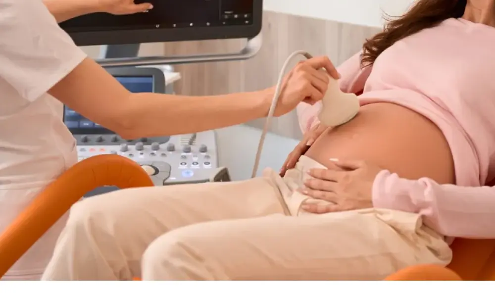
Routine anomaly scanning at 18–20 weeks identifies most cases.
Transabdominal ultrasound: Useful initial screen but less precise
Transvaginal ultrasound (TVS): This is the definitive modality and provides more accurate measurement of the placental edge-to-os distance than transabdominal scanning.
MRI: Used when placenta accreta spectrum (abnormal placental invasion) is suspected alongside previa, particularly in women with prior uterine scars
If previa is identified at the anomaly scan, repeat TVS at 32 and 36 weeks assesses whether the placental edge has migrated sufficiently clear of the os to reconsider delivery route.
Treatment Options for Placenta Previa
There is no intervention that repositions the placenta. Management is directed at maintaining maternal and fetal wellbeing until a safe gestational age for delivery is reached.
Pelvic rest: No sexual intercourse, no vaginal examinations, and no transvaginal procedures outside clinical necessity.
Activity restriction: Bed rest is not universally prescribed but activity limitation is recommended, particularly in women who have bled.
Hospitalisation: Women with active bleeding, those far from a tertiary centre, or those with complete previa entering the third trimester are often admitted from 34 weeks.
Corticosteroids: Betamethasone or dexamethasone between 24 and 34 weeks to accelerate fetal lung maturity in anticipation of preterm delivery.
Blood transfusion: For significant haemorrhage with maternal anaemia, to maintain haemodynamic stability and fetal oxygen delivery.
Tocolysis: Short-term uterine relaxants for preterm contractions concurrent with bleeding, buying time for steroid administration.
Risks & Complications for Mother and Baby
Common complications are:
Antepartum haemorrhage - repeated bleeding episodes risk progressive maternal anaemia
Postpartum haemorrhage - the lower uterine segment contracts poorly after delivery, increasing blood loss
Placenta accreta spectrum - particularly in women with prior Caesarean scars, the placenta may invade myometrium or beyond; this is the most serious associated complication and may require hysterectomy
Preterm birth - most women with symptomatic previa deliver before 37 weeks
Fetal growth restriction - placental positioning in the lower segment may compromise perfusion
Malpresentation - breech or transverse lie complicates delivery planning.
Delivery Planning in Placenta Previa
Complete and partial previa are absolute indications for Caesarean section, typically planned at 36–37 weeks in uncomplicated cases; recurrent bleeding may require earlier delivery. The surgical team cross-matches blood products in advance and anticipates significant intraoperative haemorrhage. Where accreta spectrum is suspected on MRI, a multidisciplinary team including interventional radiology, urology, and vascular surgery is assembled preoperatively. Prophylactic balloon catheters or ureteric stents may be placed before the incision at centres with appropriate expertise.
Can Placenta Previa Resolve on Its Own?
In early pregnancy, apparent low placentation frequently resolves. As the uterus grows, the lower segment elongates and the placental edge moves relatively upward. Complete previa at the anomaly scan resolves in a minority of cases; marginal previa identified early has a higher resolution rate. Previa persisting at 32 weeks is unlikely to clear sufficiently for vaginal delivery. Each case is reassessed by transvaginal ultrasound at 32 and 36 weeks before delivery planning is finalised.
Precautions & Lifestyle Tips During Pregnancy
If you have placenta previa, several tips are helpful:
Attend all scheduled ultrasound appointments particularly at the 32 and 36 week TVS are clinically decisive
Observe strict pelvic rest like avoiding sexual intercourse and internal examinations
Know the warning signs like any fresh vaginal bleeding requires immediate emergency attendance regardless of volume
Stay within reach of a tertiary obstetric facility after 28 weeks if complete previa is confirmed
Maintain iron levels - haemoglobin above 10 g/dL provides buffer against acute blood loss
Avoid straining and constipation as the Valsalva manoeuvre can precipitate bleeding
Discuss a birth plan early and understand that a planned Caesarean reduces anxiety and enables practical preparation.
FAQs
What is placenta previa and how does it affect pregnancy?
Placenta previa occurs when the placenta implants in the lower uterine segment, partially or completely covering the cervical os. It prevents vaginal delivery, creates haemorrhage risk, and requires specialist obstetric management from diagnosis through delivery.
What are the symptoms of placenta previa?
Painless, bright red vaginal bleeding in the second or third trimester. The uterus remains soft and non-tender, which distinguishes it from abruption. Many women have no bleeding; the diagnosis is made on a routine ultrasound.
What causes placenta previa?
Abnormal implantation in the lower uterine segment. Prior uterine scarring due to Caesarean section, D&C, and myomectomy is the most consistently identified predisposing factor.
Who is at higher risk of developing placenta previa?
Women with prior Caesarean sections, uterine surgery, advanced maternal age, multiparity, multiple gestation, smoking, IVF conception, or previous placenta previa.
How is placenta previa diagnosed?
Transvaginal ultrasound is the definitive investigation. It is more accurate than transabdominal scanning and safe in this context. Most cases are identified at the 18–20 week anomaly scan and reassessed at 32 and 36 weeks. MRI is added when placenta accreta is suspected.
Is placenta previa dangerous for the baby?
It carries risk of preterm delivery, fetal growth restriction, and acute fetal compromise during major haemorrhage. With specialist-managed delivery at appropriate gestation, outcomes for the baby are generally good.
What treatment is available for placenta previa?
No treatment repositions the placenta. Management is pelvic rest, activity restriction, hospitalisation when indicated, corticosteroids for fetal lung maturity, and planned Caesarean at the appropriate gestation.
Can placenta previa go away on its own?
A low-lying placenta before 20 weeks frequently resolves as the uterus grows. Complete previa at the anomaly scan has a lower resolution rate. Previa persisting at 32 weeks is unlikely to clear sufficiently for vaginal delivery.
Is normal delivery possible with placenta previa?
Complete and partial previa are absolute contraindications to vaginal delivery. Marginal previa within 2 cm of the os may be considered for vaginal delivery in carefully selected cases but Caesarean is the usual outcome.
What precautions should be taken with placenta previa?
Strict pelvic rest, no sexual intercourse, avoidance of strenuous activity, maintaining haemoglobin above 10 g/dL, immediate attendance at any bleeding episode, proximity to a tertiary obstetric unit in the third trimester, and consistent follow-up ultrasound attendance.